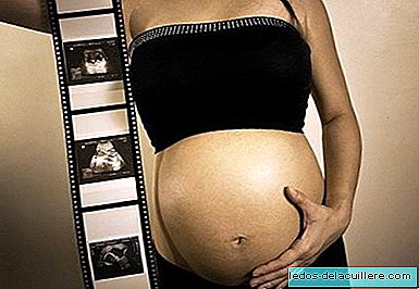
Ultrasound has marked a before and after in the control of pregnancy. It is the very important prenatal test because of the amount of information it is able to offer. Thanks to ultrasound in pregnancy The intrauterine development of the baby can be evaluated, so it is vital from the medical point of view, but it also allows parents to see their child and hear their heartbeats before birth, being also very valuable emotionally.
It is also known as ultrasonography or echo sonography and it consists of a non-invasive technique that allows, through ultrasound waves, to see images of organs and structures inside the body, and in the case of pregnant women, to examine the fetus inside the mother's womb.
In a test that began to be used in the 70s, experiencing a breakthrough to this day. The images are obtained by a transducer that sends the sound waves and a gel that serves as a transmitter.
Ultrasound in pregnancy is mainly used to study the growth and well-being of the baby inside the womb. It allows, among other things, to detect malformations, evaluate the anatomical development of the fetus, its growth, its position, calculate its gestational age, hear its beats, observe the state of the placenta, amniotic fluid, estimate the baby's weight and detect abnormalities or pathologies of pregnancy.

2D or two-dimensional ultrasound is the most widespread, but in recent years we have also known 3D ultrasound that offers us three-dimensional images with a volume aspect and the so-called 4D, which also adds the complement of seeing the baby moving in time real.
In the future it may be common to have ultrasound at home, for example through mobile phones, but even so, ultrasounds done by a specialist capable of giving us a diagnosis of how our baby grows will be necessary.
It is a simple, harmless and painless technique. It does not involve radiation or exposure, so it is a safe practice for both pregnant and baby. No adverse effects have been demonstrated for either the baby or the mother, although it is recommended not to abuse its use and only perform those that the doctor considers necessary.
They are usually performed three routine ultrasound exams throughout pregnancy. One in each quarter. Sometimes one is also done in the first weeks of pregnancy, which is usually vaginal, to confirm pregnancy. Until about the 8th week they are done in this way to have an early diagnosis of the fetus and be able to record their beats.
The other three are abdominal. It is possible that if you carry your pregnancy through private medicine, you will have an ultrasound scan at each monthly check to see how the pregnancy develops.
The first ultrasound

For some, the first ultrasound will be the confirmation of pregnancy in the first weeks of pregnancy. The size of the fetus is measured, the amount of embryos and the embryo implantation site are observed to rule out a possible ectopic pregnancy.
Otherwise, the first one will surely be done in the 12th week of gestation. It allows to determine if there are one, two or more embryos, to know the position in the uterus, to listen to the heartbeat and to measure the fetus to determine if it fits the real time of gestation.
The measurement of the nuchal fold is also performed, the most sensitive and specific early ultrasound marker suspected of Trismomy 21 or Down Syndrome.
The second ultrasound

The second ultrasound scan is performed in the 20th week of gestation. It allows to confirm that the fetal growth is correct, assess the heartbeat, the movements of the fetus, its morphology and observe its internal organs.
It is checked, although not with 100 percent reliability, if there is any malformation so it is usually a more thorough and detailed exploration. The level of amniotic fluid, the functioning of the placenta and the umbilical cord are also controlled.
It is very likely that this ultrasound can know the sex of the baby, as long as your position allows.
The third ultrasound

The third ultrasound is usually performed between week 33 and 35 of gestation. It is already the last ultrasound before the birth of the baby, so it mainly serves to obtain information about how the birth will develop.
In addition to assessing the condition of the fetus, its growth, heartbeat and movements, it also serves to know its position inside the uterus: if it is already upside down, if it is embedded in the pelvis, if it is not, if it is buttocks, etc.
As the baby has grown quite at this point, only parts of his body can be seen on ultrasound.
Doppler ultrasound

It is not a routine ultrasound, but in some cases an ultrasound is usually done with Doppler technique. It is used to measure and evaluate the flow of blood that circulates through the arteries and cavities and valves of the baby's heart.
It can be seen blood flow in color, allowing early detection of any anomaly related to the circulatory flow and the functioning of the heart.
It also allows to evaluate if the blood circulation of the umbilical cord is adequate.
It is indicated to control pregnancies in which the mother has hypertension, diabetes, problems with the placenta or umbilical cord, growth retardation, fetal distress, or cardiac malformations of the baby.
4D ultrasound

4D ultrasound (or 3D plus real-time movement) is a most fashionable technique lately. It does not supplant traditional ultrasound, but complements it. It is an ultrasound that is done on a private level, so there are several things to consider before hiring it.
From the point of view of detection of fetal anomalies, it does not add value to the previous ones at the level of functioning of organs, morphology, growth of the baby, etc. It may be more valuable to detect, for example, skin problems or deformities such as cleft lip or palatal fissure.
It is still a prenatal diagnostic test, but it is transcendent more than anything since the emotional point of view, as it allows parents to know their baby's face. The images are quite clear but it is not a photo, it only allows us to get an idea of its features.
4D ultrasounds can be performed at any time during pregnancy although the best images are obtained between weeks 24 and 30 because the conditions are the most appropriate. The proportion of amniotic fluid and the intermediate size of the baby allow you to visualize it better.
Photos | Anthony Muñoz, myllissa, seamusiv, jula julz, julia julz, Daquella way, Mrs Finger on Flickr In Babies and more | Prenatal tests: ultrasound