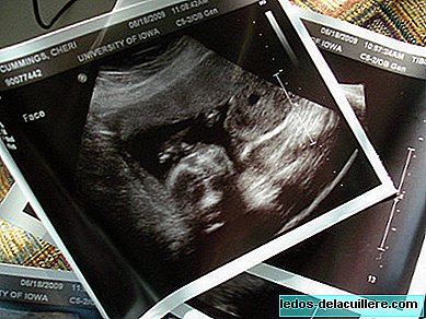
During the 32nd and 36th week of pregnancy, what will normally be the last ultrasound of the pregnancy, known as ultrasound of the third trimester. It will check the general condition of the baby and its surroundings.
This ecogrfía does not require such a high degree of specialization nor a high quality equipment, such as that of week 20, although you will get very valuable information about the state of the baby and its evolution for the delivery.
Since the baby is already in an advanced state of development, he has less space to move and has accumulated enough calcium in his bones, all this causes that the ultrasound does not work at 100% transfer well and therefore The information we can get is not as detailed as in the previous ones. That is why this echo is aimed more at collecting information on the position of the baby, the umbilical cord and the placenta in order to prepare for the delivery.
Remember that once we reach the 37th week of pregnancy, the pregnancy is considered to have come to an end, so when this ultrasound is done The end is already very close.
What parameters are studied?
He approximate baby weight, to know if it is suitable for the time in which we are and rule out growth problems.
Check the position of the placenta, as well as its status. It is important to know this fact, since a previous placenta can cause complications in a vaginal delivery and have to perform a C-section. It also suits check that the placenta does not show excessive aging, what is called old placenta or hypermadura, if so, it will be the doctor who assesses if it is necessary to start any treatment to increase blood flow.
Study umbilical cord position and examine the functioning of the umbilical artery to verify that the baby's oxygenation is correct.
Measure amniotic fluid levels and verify that they are suitable for these weeks.
Rate the biophysical profile of the baby: your heart rate, your respiratory and body movements and fetal tone.
Detection of late abnormalities in the baby, Although, as we have said, it is not the most accurate ultrasound that will be done if some anomalies can be detected, usually in internal organs.
Cervical length The length of the cervix is a factor that helps predict the possibility of premature delivery. During pregnancy it measures about 3-4 centimeters. When labor begins, in a first phase, the neck shortens until it disappears, from here, the dilation begins. In some cases, the neck may shorten prematurely, increasing the risk of preterm birth.


