I don't know how you are interpreting ultrasound of babies, but there are many fathers and mothers who after witnessing one of their future baby come out saying that they have not been able to see practically anything. Yes, they saw things that moved and sometimes seemed to see something, but in general they had not deciphered very well "the message".
For a few years, when high definition arrives at ultrasound scanners, the interpretation is simpler; although it is true that there are times when it is still difficult to know what we are seeing. Well, it is likely that this feeling of the parents will come to an end, if the magnetic resonance ultrasound scans and become habitual. The differences are quite evident: a video shows what the 'ultrasound' will look like in the future.
The iFind project
Created in London with a budget of 10 million pounds, the iFind project's mission is to achieve the technology necessary to perform routine ultrasounds using magnetic technology and radio waves.
In view of what can be seen in this video, they have achieved it:
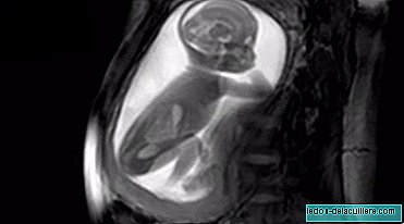
The material they get is so detailed that you can see the baby turning the neck, playing with the cord, jumping, meeting, etc. In the case of the video, the fetus is 20 weeks old and that is why the whole body is seen.
And this is the most incredible: by means of algorithms, the ultrasound machine is able to capture the image of the entire uterus and even the mother's body, in high quality. Nothing to see an arm, now a leg, now the head, now "I don't know what this is", etc. In a single image the baby is seen completely, so that you can have a very clear idea of what you are doing at each moment.
An ultrasound that will also be diagnostic
Thanks to the method of image capture, not only can the exterior of the baby be seen, but also the interior, with great definition. In this way, even babies who move a lot in the womb can be recorded to analyze the images carefully, and assess whether everything is developing normally.
The video, just 24 seconds old, has been shared on the ChannelMum.com baby portal, to be shared with all pregnant mothers in the world.
He Dr. David Lloyd, a clinical researcher at Kings College London, who has been part of the project, explains it in DailyMail:
You can see the internal structures of the body, regardless of whether there is bone, muscle or fat in the path; and in some cases it can give us even more detailed images than an ultrasound. Importantly, it is also one of the few imaging techniques that are safe during pregnancy.
A breakthrough for when babies move a lot
As I have just explained, the novelty of the system is not only the definition and the ability to cover a large field of vision, but also to be able to obtain, in a stable way, images of babies that move more than desirable to see them carefully .
There are many ultrasounds in which the baby is so restless that it is barely visible, while with this system stable and valid results can be obtained, very useful to make possible diagnoses before and, consequently, to be able to act also before, if some type of medical intervention is needed.
Now we just need this technology to be designed - they are devising an automatic image capture system that works with four sensors from different angles - and from that moment the cost-benefit of the advance is assessed, in case hospitals consider it appropriate to start to use it